|
The Application of Osteopathic Principles and Practices in Managing Upper Respiratory Track Infections in a Pediatric Population
Vadhya Elivert, DO
Anne Marie Zeller, DO
Michael P. Rowane, MS, DO, FAAFP, FAAO
_________
Osteopathic Approach to Lower Respiratory
Tract Infections in Pediatric Patients
Deena L. Varber, BSN, RN
Vadhya Elivert, DO
Michael P. Rowane, MS, DO, FAAFP, FAAO
_________
The Application of Osteopathic Principles
and Practices in Managing Upper Respiratory Track Infections in a Pediatric Population
Vadhya Elivert, DO
Traditional Intern, Department of Medical Education
University Hospitals Regional Hospitals
Richmond Heights, OH
Anne Marie Zeller, DO
Family Medicine Intern,
Department of Medical Education
University Hospitals Regional Hospitals
Richmond Heights, OH
Michael P Rowane, DO, MS, FAAFP, FAAO
Director of Medical Education
University Hospitals Regional Hospitals
Richmond Heights, OH
Abstract:
Pediatric Upper respiratory infections are common clinical presentations in both the emergent setting and primary care office. “The average incidence of the common cold in preschool children is 5 to 7 per year, but 10% to 15 % of children will have at least 12 infections per year.”(1) Many physicians are prescribing antibiotics for what are self limiting viral infections because of the recurrence and symptomatic discomfort. Common clinical findings of otitis media and sinusitis are discussed. There are studies that show that the use of osteopathic manipulation have helped decrease the incidences of upper respiratory infections in the pediatric population, lower the recurrence rate, increase the immune response as well as provide symptomatic relief. This article describes a basic algorithm to reflect the current medical literature that may prove helpful in the treatment of the most common presentations of upper respiratory infections.
Introduction
Upper respiratory tract infections account for a majority of pediatric clinic visits, often followed by or accompanied with the complication of otitis media, also referred to as: the common cold and ear infection. It accounts for approximately 22 million days of absence from school and many days of work absences of parents who must care for their sick children.(2) Upper respiratory infections (URI) include a vast number of diagnosed ailments, but most commonly clinically presented in the pediatric population are sinusitis, pharyngitis, common cold, and otitis media. A majority of URIs are of viral etiology while approximately 20-30 % do not have a proven viral cause,(2) which puts them in one of two categories: 1) undetected viral etiology or 2) bacterial etiology.
There are various factors that contribute to the incidences of URIs in the pediatric population. One of them are time of year. Although URIs can be contracted year round, the fall through winter months are the peak times for contraction. Day-care attendance is another major risk factor for the number of URI incidences. This may explain why infancy through 4 years old have the highest mean annual incidence of respiratory illness per person.(2) Incidences of URIs decrease as age increases. “The average incidence of the common cold in preschool children is 5 to 7 per year, but 10% to 15 % of children will have at least 12 infections per year.”(1)

Pathophysiology
The upper respiratory tract consists of the airways from the nostrils to the vocal cords, larynx, paranasal sinuses and middle ear. When an irritant or invader infects a host, the body’s inflammatory response such as rubor, calor, dolor, and tumor, all take effect. Inflammatory mediators and complement factors are released by the resident macrophages and act as a positive chemotaxis attracting other immune cells in to respond and migrate to the area. Other chemicals released such as bradykinin, histamine, serotonin and leukotrienes sensitize pain receptors in the area resulting in a dilation of blood vessels to the affected area in order to allow more immune cells into the area to fight the foreign body.
While this is occurring, the adaptive immune system is mobilizing and as the number of mobilized and fighting cells increases, the surrounding lymphatic tissue also swells and increases in size. This presents in the red, swollen, and sore tissue of pharyngitis. The tender, congested sinuses of sinusitis, the bulging erythmatous, swollen tympanic membrane of otitis media, the palpable and tender cervical lymph nodes of the cervical area and the general symptoms of congestion and malaise are associated with an upper respiratory tract infection.
Otitis media is one of the most common infections in children and one of the top three reasons for pediatric office visits for ages 0 to12.(3) According to one study, more than 60% of symptomatic URI in young children were complicated by otitis media.(4) Otitis media is an inflammation of the air filled middle ear space. This inflammation can be either by viral or bacterial infection or of Eustachian tube dysfunction; often it is a combination of both an infectious process and anatomical variation found in the pediatric population. The inflammatory process resulting from infection results in swelling of the tissues within the surrounding area including the Eustachian tube, resulting in a decrease in drainage of fluid accumulation in the middle ear. This contributes to the symptoms associated with an ear infection.
Common History and Physical Examination Findings
Diagnosing upper respiratory tract infections in the pediatric population in the osteopathic setting requires a combination of history taking and physical examination and palpatory skills. For the pediatric population, much of the history will be given by the parent or guardian of the patient, as the pediatric age group with the highest incidence of URIs is ages six and under. Older age groups may contribute more to the history as verbal communication skills develop more.
In general, history of symptoms may include, rhinorrhea, head congestion, cough (both productive and non productive), sneezing, nasal obstruction, sore throat, “scratchy” throat, fever, hoarseness, headache malaise, and lethargy. Clinical presentation will vary depending on age, presentation day of child within course of illness, particular region affected by URI and personality, to name a few.
Physical exam findings may include, tender facial sinuses, erythamatous and/or edematous oropharynx, nasal turbinates, tympanic membrane, and external acoustic meatus, posterior/anterior cervical and submandibular lymph nodes.
Some external structural exam findings may include tenderness below the pinna of the affected ear, swollen cervical lymph nodes on the ipsilateral side of the affected ear. If visualization inside the ear canal can be obtained, observable structural changes in structure may include fluid buildup behind the tympanic membrane, a bulging tympanic membrane, inflammatory changes surrounding the tympanic membrane or an opaque appearance to the ear drum with loss of the ‘cone of light” typically seen in a healthy ear drum.
Osteopathic findings may include: palpable enlarged anterior/posterior cervical lymph nodes, somatic dysfunctions along the vertebrae from C1 thru T4, palpable anterior and posterior corresponding Chapman points, restricted cervico-thoracic inlet, tissue texture changes around the head and cervical area, rib dysfunctions, etc.
Osteopathic Evidence-Based Literature
Pediatric upper respiratory infections are generally self limiting and studies have shown that osteopathic manipulation does help facilitate recovery and prevent future infections. There are positive findings when reviewing literature to determine the effectiveness of osteopathic manipulative treatment of upper respiratory infections in the pediatric patient population.
“The average incidence of the common cold in preschool children is five to seven per year, but 10% to 15% of children will have at least 12 infections per year.”(1) The incidences of URIs decrease as age increases. A small pilot cohort study by Degenhardt and Kuchera (2006), aimed at studying the effectiveness in reducing the morbidity of otitis media with OMT showed that with just three weeks of manipulative treatment, 62.5% of the patient subjects had no recurrent symptoms of otitis media.(5) Saggio et al (2011) showed a positive correlation between the effect of OMT on serum IgA levels. High IgA levels in humans have been connected to fewer incidences of upper respiratory tract infections.(6) The goal is to help the body heal itself by returning it to its most stable state through normalization of biomechanical structure and facial tissues.
An early study compared the complication rate of URIs after mainly antibiotic treatment verses symptomatic treatment and osteopathic manipulative treatment. The results of the study showed that when comparing these different treatment modalities, manipulative treatment had the lowest percentage rate of complications at 5%.(7) If manipulative treatment can help reduce the recurrences of URIs that would result in fewer missed school days, work day, symptomatic relief, improved immunity and decreased risk of complications resulting from recurrent URIs.
Osteopathic Concepts Applied to Treatment of URIs
Results from various studies presented in previous section showed the usefulness of manipulation and it’s application within the pediatric population. This literature has assisted the authors in developing a general osteopathic treatment protocol for the management of upper respiratory infections
There are four basic conceptual ideas that any clinician must keep in mind that will cover the general osteopathic management of URIs with pediatric patients:
1. Release of the thoracic inlet, and other related physiologic facial diaphragms
2. Treatment of the cervico-thoracic dysfuctions,
3. Sinus and lymphatic drainage
4. Treatment of the corresponding Chapman reflex points.(8)
These concepts will cover the most common presentations and ailments involving upper respiratory infections in the pediatric population and can easily be applied and adapted in any age group. The protocol described here contain some techniques that parents can be taught and replicate to further the benefits beyond the office visit.
Osteopathic Approach to Treating Upper Respiratory Tract Infections
The authors have established a generalized approach to utilizing Osteopathic Manipulative Treatment [OMT] as a Management tool in treating Upper Respiratory Tract [URI] infections in a pediatric population. The following steps may be helpful as an adjunct in treating patient with a URI, sinusitis, pharyngitis and otitis media.
- Step 1 of this approach is diagnosis and treatment of cervicothoracic junction.
- Step 2 is treatment of suboccipital region.
- Step 3 is to evaluate and treat cervical and upper-mid thoracic spine.
- Step 4 is generalized treatment of sinuses.
- Step 5 is treatment of Eustachian tube dysfunction.
- Step 6 is treatment of Chapman points
STEP 1: Diagnosis and Treatment of Cervicothoracic junction/Thoracic Inlet
Osteopathic Concepts Utilized
Obvious structural findings of the upper respiratory infection will include palpable lymph nodes. Osteopathic treatment of a URI should initially focus on the draining of the lymphatic system to aid in the healing process. Much of the discomfort that patients experience stems from restrictions in the draining of the lymphatic system especially from the head and neck area in upper respiratory infections.
The thoracic duct is the largest lymphatic vessel in the body, it drains into the venous system around the junction of the left subclavian and left brachiocephalic vein. All lymph from the left side of the head, neck, left arm, thorax lower body and thoracic viscera drain into the thoracic duct. The right side of the head, neck, arm and chest empty into the right lymphatic duct. Both ducts pass through the fascial plane of the thoracic inlet [cervicothoracic region], so dysfunction in this area can easily obstruct lymphatic flow.
Anatomically, the thoracic inlet includes the first ribs, the first thoracic vertebra and the manubrium. Clinically, however it includes the first two ribs, the first four thoracic vertebra and the manubrium of the sternum, that in conjunction with fascia from the scalenus and longus coli muscles and a the function thoracic inlet diaphragm is formed.(9)
Structural Examination Findings
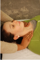
Structural exam finding may include limited range of motion with side bending and rotational aspect of the clavicles , upper thoracic and cervical area, tight musculature of the neck and upper back, corresponding Chapman points on the anterior of the patient and tender swollen upper lymphatics.
 |
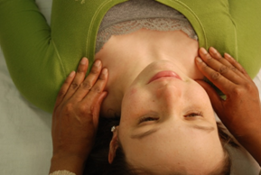 |
Figure 1A Diagnosis of the Cervicothoracic junction/Thoracic Inlet |
Figure 1B Diagnosis of Clavicular
restrictions that affect the Cervicothoracic
junction/Thoracic Inlet |
- The patient is placed in the supine position.
- The clinician positions themselves at the head of the patient.
- Also rotational motion can be assessed by positioning the hands palm side down, cupping ipsilateral shoulders between the thumb and the rest of the four digits. The thumb would be placed on the posterior portion of the patients shoulder while the other four digits would be placed on the anterior aspect of the patient’s shoulder. As if the clinician were “holding onto a horizontal staff.”
- A downward force towards the table should be applied to both shoulders, followed by an upward force to determine where the motion of ease exists.
- Hand positioning to assess side bending can done by two methods; 1) Alternatively pushing down caudally through the shoulders to see which side is restricted in motion or 2) The clinician assumes the “staff holding” position as previously described and administers the alternating caudal ( relative to the supine patient) force to elicit side bending motion. The side with the least amount of motion is the side of the side bending dysfunction.
OMT Technique(s):
Direct and Indirect Myofascial Release Technique for the Thoracic Inlet
Osteopathic Concept Utilized
Dysfunction of the thoracic inlet can be treated with myofacial release both direct and indirectly. The goal of either method is to cause a release within the facial structures in the diaphragm. The basic idea of treatment is to move the tissue in the combined planes of rotation and side bending either directly towards the barrier/restrictions and hold position until a release is felt, or towards the direction of ease indirect till a release of the fascial tissue is felt.(10, 11)
Figure 2. Direct and Indirect Myofascial Release Technique for the Thoracic Inlet
Description: Indirect
- Patient is in supine position.
- Clinician places hands in “staff holding“ position.
- Clinician moves inlet in both rotational motion of ease and
side bending motion of ease.
- Position is held until a release if felt or tissue softens.
Step 2: Treatment of Suboccipital region
Osteopathic Concepts Utilized
The occipital fascial is another region in which lymphatics pass. Because of the muscular interaction with the occipital fascia, drainage of the sinuses as well as some lymphatic nodes may be impeded if the occipital fascia and its adjoining musculature are restricted. Release of the occipital fascia will help with drainage and lymph flow.
Structural Examination Findings:
Structural exam findings may include tissue texture changes of the occipital fascia along with tissue changes of themusculature of the cervical area, tenderness upon palpation, along with some corresponding Chapman points in the suboccipital area.
OMT Technique(s):
Suboccipial Tension Release Inlet |
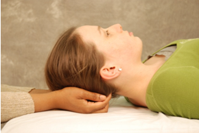 |
Figure 3 Suboccipial Tension Release Inlet
Description |
- The patient is placed in the supine position.
- The clinician positions themselves at the head of the patient.
- With palms facing upwards towards the ceiling, the clinician places their hands underneath the patient’s head cupping the suboccipital region.
- Using finger pads of the second through fifth digits the clinician applies a gentle but firm upwards and into the region where the trapezius muscle and occipital fascia meet.
- This position is held for approximately 10-15 seconds, then is released.
- This process can be repeated for approximately up to one minute.
Step 3: Evaluate and treat sites of somatic dysfunction in the cervical and upper-mid thoracic spine.
Osteopathic Concepts Utilized
The autonomic innervations for the head and neck arise from both the sympathetic and parasympathetic system. The sympathetic innervations originate from the nuclei of the T1-T4 segments of the spinal cord and their ganglionic neurons are located in the cervical sympathetic ganglia of the neck. Somatic dysfunction of the cervical or thoracic vertebrae can result in over or under stimulation of either the sympathetic or parasympathetic system, that can exacerbate the symptoms of an upper respiratory infection. Treatment of these vertebral dysfunctions will help alleviate certain symptoms and normalize and regulate the body to promote healing.(9)
Recommended OMT Techniques:
Physician may utilize varying OMT techniques to treat sites of somatic dysfunction found in the cervical and thoracic regions. OMT techniques that are easily tolerated and researched in a pediatric population include soft tissue techniques, muscle energy, myofascial release and strain/counterstrain. The application of these techniques is not the focus of this paper, but can easily referenced in standard osteopathic texts, including Foundations of Osteopathic Medicine.(9)
Step 4: Generalized Treatment of Sinuses:
Trigeminal Stimulation [Supra-Orbital, Infra-Orbital and Sub-Mental]
Effleurage of the Frontal and Maxillary Sinuses
Osteopathic Concepts Utilized
In the pediatric population, the sinuses develop and mature at different stages in life ranging from in-utero till early adulthood. The maxillary and ethmoid sinuses begin development around the third to fourth month of gestation and by birth are only a few millimeters in size, they reach full size by adolescence. The sphenoid sinuses begin formation around age two and reach their full size around adolescence. The fontal sinuses are the last to develop, beginning at approximately age four and reaching full size in early adulthood.
In a healthy patient, the mucus secreted is generally thin and clear, however in cases where irritation, infection, or inflammation occur, these secretions can become very thick and viscous. The tissue within the sinuses themselves become very edematous, and ciliary movement slows down, resulting in an accumulation of secretions causing a buildup of pressure with no outlet within the sinuses. This results in the feeling of pressure or pain associated with sinusitis. In cases of sinusitis within the pediatric population, several techniques using myofacial massage in the facial area around the sinuses help stimulate drainage of the surrounding tissues to relieve pressure within the sinuses. Within these fascia planes are the supraorbital, infraorbital, and mental nerves that can be stimulated in these areas resulting in a thinning of the secretions, and constriction of the vessels allowing for increase in sinus drainage.
Structural Examination Findings:
Structural exam findings may include, “boggy”, slightly edematous tissue texture changes around the maxillary sinus areas and/or tenderness upon palpation around the frontal sinus area and maxillary sinuses.
OMT Technique: Trigeminal Nerve Stimulation [Supra-Orbital, Infra-Orbital and Mental] (11) |
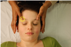 |
| Figure 4 Trigeminal Stimulation: Supra-Orbital |
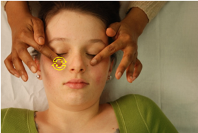 |
Figure 5 Trigeminal Stimulation: Infra-Orbital |
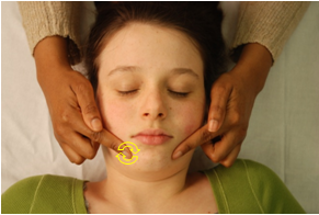 |
Figure 6 Trigeminal Stimulation: Mental |
Description:
- The patient is placed in the supine position.
- The clinician positions themselves at the head of the patient.
- Seated at the patient’s head, the clinician may use either combination of their thumb, index finger or both index and middle finger together and place the pads of either options flat and gently upon the first position of the supra orbital notch. (Location, the clinician will feel for a notch in the bone structure a few millimeters superior and medial to the orbits.)
- With the pads of their fingers, the clinician will gently make small circular motions going in either a clock wise or counter clockwise direction.
- Repeat this action for one to two minutes, as tolerated by the patient, due to this technique causing uncomfortable sensations.
- Next the clinician will repeat these steps at the second position, at the infra orbital notch. (This notch in bone structure can be found inferiorly and midway of the orbits.)
- The third position to repeat this technique is in the mental foramen. (Location is found caudally from the supra and infra orbital foramen, along the mandible of the patient.)
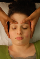
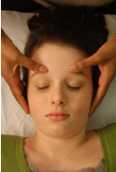
OMT Technique: Effleurage of the Frontal Sinuses and Effleurage of the Maxillary Sinuses (10,11) |
 |
 |
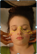 |
Figure 7A |
Figure 7B |
Figure 7C |
Figure 7A-C
Effleurage of the Frontal Sinuses |
Description: Frontal
- The patient is placed in the supine position.
- The clinician positions themselves at the head of the patient.
- The clinician may use either combination of their thumb, index finger or both index and middle finger together. Place finger pads gentle upon the first position (Figure 7A) beginning medially and superior to brow ridge.
- The clinician will then make slow, stroke-like sweeping movements laterally following the supra orbital ridge until they reach the area of the pterion.
- This action is repeated for one to two minutes.
 |
 |
 |
|
Figure 8B |
|
 |
 |
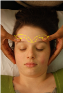 |
Figure 8D |
|
Figure 8F
|
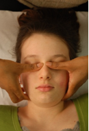
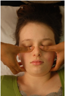
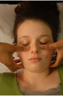
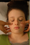
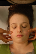
Figure 8A-F Effleurage of the Maxillary Sinuses |
Description: Maxillary Effleurage
- The patient is placed in the supine position.
- The clinician positions themselves at the head of patient.
- The clinician may use either combination of their thumb, index finger or both index and middle finger together. Place finger pads gently upon the patients face, beginning with each finger laterally on the bridge of the nose.
- The clinician will then make slow, stroke-like sweeping movements laterally following the zygomatic process of the maxilla.
- This action is repeated for one to two minutes.
Step 5: Galbreath and Auricular Drainage techniques to treat Eustachian Tube Dysfunction (10,11)
Osteopathic Concept Utilized
Eustachian tube dysfuctions are related to intrinsic anatomical differences secondary to the incomplete development of the pediatric cranial structures. They include the differences in diameter size from a child to an adult, size and development of the muscles that open the tube, to the angle of the tube itself to the middle ear. The middle ear is connected to nasopharynx by the eustachian tube. The eustachian tube traverses from the temporal bone to the nasopharynx at only a ten degree angle in children compared to a 45 degree angle in an adult. This developmental difference promotes fluid and pooling, increasing the opportunity of infection to develop. The galbreath technique is used to assist in opening the eustachian tube in order to help with drainage, while auricular drainage technique is used to help release any dysfunction or lymphatic congestion in the ear region.
OMT Technique: Galbreath Technique |
 |
 |
Figure 9A |
Figure 9B |
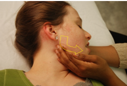
Figure 9A-B Galbreath Technique |
Description
- The patient is in supine position with their head rotated with affected ear facing upwards.
- The clinician seated facing the patient at the head of the patient, places their most cephalad hand palm side up underneath the patient's head on the contralateral side of treatment to elevate slightly, support and stabilize the patient’s head.
- Next, the clinician's most caudal hand is placed along the mandible of the patient with the second and/or third finger tips gently cupping the angle of the mandible with the rest of the digits along the body of the mandible.
- In older pediatric patients with more advanced communication skills, the clinician gives the patient instructions to open their mouth slightly.
- The clinician then gently draws the mandible slightly forward, towards the clinician, and towards the midline of the patient or medially towards the table.
- This position is held for approximately ten seconds and then released. This process is repeated rhythmically for 30 seconds to two minutes.
OMT Technique: Auricular Drainage technique |
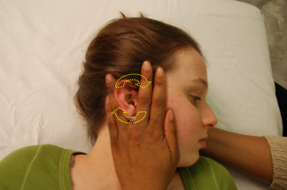 |
Figure 10 Auricular Drainage |
Description
- The patient is in the supine position with their head with affected ear facing upwards.
- Place their most cephalad hand palm-side up underneath the patients head on the contra lateral side of treatment to elevate slightly, support and stabilize the patient’s head.
- The clinician then groups their second and third digits together and fourth and fifth digits creating a “V” space in the center of their grouped digits of their most caudal hand( in relation to the patient).
- Palm-side down towards the table, the clinician places their hand flat against the side of the patient’s head with the tips of their fingers pointing in the cephalad direction of the patient and the patient’s ear within the “V” space created or between their third and fourth digits.
- The clinician then, with very gentle pressure on the patient’s head towards the table, makes gentle circular motions in either a clockwise or a counterclockwise motion for approx 15 seconds then reverses the direction of motion for another 15 seconds. This can be repeated for approximately one to two minutes.
Step 6: Assess and Treat Using Chapman’s Points for Head and Neck
Osteopathic Concepts Utilized
Chapman reflex points are somatic finding palpated, that correspond to visceral dysfunctions occurring within the body. The osteopathic profession defines Chapman reflexes as “a system of reflex points that present as predictable anterior and posterior fascial tissue texture abnormalities assumed to be reflections of visceral dysfunction or pathology.”(9) This includes lymphatic, neuro, endocrine and autonomic responses to injury, disease, as predictable palpable somatic points of visceral dysfunctions.
The approach to diagnosing Chapman reflex points may vary depending on their location. Variations from a light palpating motion to firm circular motions may be the difference in feeling certain anterior points to certain posterior points along the thoracic vertebrae. For a Chapman reflex to be positive, both the anterior and its corresponding posterior point must be present. Treating Chapman points requires identification of the point and direct myofacial stimulation to the point itself.(8)
Structural Examination Findings:
The Chapman points associated with upper respiratory infections might present as edematous, ropy, “shotty” or “fibrospongy” points, a few millimeters in diameter, found around the mid clavicular, parasternal area, the occipital region and upper para cervical regions.
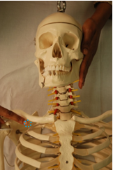
Chapman Point- Sinuses / Nasal Sinuses |
 |
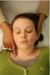 |
Figure 11A |
Figure 11B |
Figure 11A-B Anterior Chapman Point- Nasal Sinuses |
 |
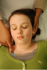 |
Figure 12A |
Figure 12B |
Figure 12A-B Anterior Chapman Point- Sinuses |
Description: Nasal sinus
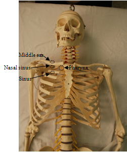
Anterior points: are located on the inferior border of the proximal third portion of the clavicle; bilaterally.
Posterior points: are located midway between the mastoid process and the inion; bilaterally.
Sinus
Anterior points: are located straight down from the nasal sinus points on the superior border of the 2nd rib; bilaterally.
Posterior points: are located on the C2 articular pillars.
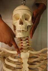
Chapman Point-Pharynx |
 |
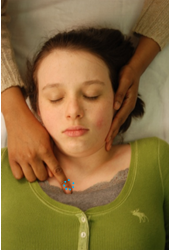 |
Figure 13A |
Figure 13B |
Figure 13A-B Anterior Chapman Point- Pharynx |
Description
Anterior points: are located on the junction between the first rib and the manubrium, below and slightly lateral to the sternoclavicular junction; bilaterally.
Posterior points: are located on the C2 articular pillars.
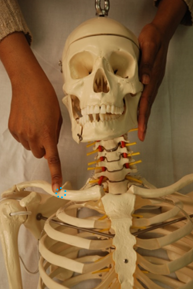
Chapman Point-Middle Ear |
 |
 |
Figure 14A |
Figure 14B |
Figure 14A-B. Anterior Chapman Point- Middle Ear |
Description
Anterior Points: are located on the superior edge of the proximal third portion of the clavicle; bilaterally.
Posterior points: are located on the posterior rami of C1.
OMT Technique(s): Fundamental Approach
to Treatment of Chapman Points (10) |
 |
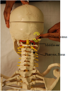 |
Figure 15 “Anterior Chapman Points” |
Figure 16 “Posterior Chapman Points” |
Description:
- The patient is placed in the supine position.
- The clinician positions themself at the head of the patient.
- Seated at the patient’s head, the clinician may use either combination of his index finger or both index and middle finger together and place the finger pads of either options flat and gently upon the corresponding positive reflex points.
- The clinician will then apply gentle pressure into the point and make slow firm circular movements on the area. This is to be done for 20-60 seconds.
- The clinician will then reassess for presence of reflex points.
- These points can be uncomfortable so treat as patient can tolerate.
Discussion / Conclusion:
This article is a basic introduction to a generalized protocol to treating pediatric upper respiratory infections. Evidence-based studies reinforce each other in the benefits of osteopathic manipulative treatment. Although there are several small studies showing enormous potential, more research needs to done in this field. There are many advantages to the use of OMT within the pediatric population as an adjunct to pharmacology and other supportive therapies.
References:
- Turner, Ronald B. "Epidemiology, Pathogenesis, and Treatment of the Common Cold." Annals of Allergy, Asthma & Immunology 78.6 (1997): 531-40. Print.
- Heikkinen, Terho, and Asko Jarvinen. "The Common Cold." The Lancet 361 (2003): 51-59. Print.
- Lund, Gregg, DO, MS, and Jane E. Carreiro, DO. "Characteristics of Pediatric Patients Seen in Medical School-Based Osteopathic Manipulative Medicine Clinics." Journal of the American Osteopathic Association 110.7 (2010): 376-80. Print.
- Chonmaitree, Tasnee, Krystal Revai, James J. Grady, Audra Clos, Janak A. Patel, Sangeeta Nair, Jiang Fan, and Kelly J. Henrickson. "Viral Upper Respiratory Tract Infection and Otitis Media Complication in Young Children." Clinical Infectious Diseases 46.6 (2008): 815-23. Print.
- Degenhardt, Brian F., DO, and Michael L. Kuchera, DO. "Osteopathic Evaluation and Manipulative Treatment in Reducing the Morbidity of Otitis Media: A Pilot Study." Journal of the American Osteopathic Association 106.6 (2006): 327-34. Print.
- Saggio, Gregory, DO, Salvatore Docimo, DO, Jennifer Pilc, DO, Jennifer Norton, DO, RN, and Wolfgang Gilliar, DO. "Impact of Osteopathic Manipulative Treatment on Secretory Immunoglobulin A Levels in a Stressed Population." Journal of the American Osteopathic Association 111.3 (2011): 143-47. Web
- Purse, F. Monro, DO. "Manipulative Therapy of Upper Respiratory Infections in Children." Journal of the American Osteopathic Association 65 (1966): 964-72. Print.
- Kuchera, ML, Kuchera, WA.Osteopathic Considerations in HEENT Disorders. Greyden Press. 2012.
- Chila, Anthony G. Foundations of Osteopathic Medicine. Philadelphia: Wolters Kluwer Health/Lippincott Williams & Wilkins, 2011. Print.
- DiGiovanna, Eileen L., Stanley Schiowitz, and Dennis J. Dowling. An Osteopathic Approach to Diagnosis and Treatment. Philadelphia: Lippincott Williams and Wilkins, 2005. Print.
- Nicholas, Alexander S., and Evan A. Nicholas. Atlas of Osteopathic Techniques. Philadelphia: Wolters Kluwer Health/Lippincott Williams & Wilkins, 2008. Print.
- Hayes, Natalie M., DO, and Todd A. Bezilla, DO. "Incidence of Iatrogenesis Associated With Osteopathic Manipulative Treatment of Pediatric Patients." Journal of the American Osteopathic Association 106.10 (2006): 605-08. Print.
- Kline, Charles A., DO. "Osteopathic Manipulative Therapy, Antibiotics, and Supportive Therapy in Respiratory Infections in Children: Comparative Study." Journal of the American Osteopathic Association 65 (1965): 278-81. Print.
- Schmidt, Ida C., FACGP, FAO. "Osteopathic Manipulative Therapy as a Primary Factor in the Management of Upper, Middle, and Pararespiratory Infections." Journal of the American Osteopathic Association 81.6 (1982): 382-88. Print.
- Vesa, Sirpa, MD, Marjaana Kleemola, MD, Soile Blomqvist, MSC, Aino Takala, MD, Terhi Kilpi, MD, and Tapani Hovi, MD. "Epidemiology of Documented Viral Respiratory Infections and Acute Otitis Media in a Cohort of Children Followed from Two to Twenty-four Months of Age." The Pediatric Infectious Disease Journal 20.6 (2001): 574-81. Print.
_____________________________
Osteopathic Approach to Lower Respiratory Tract Infections in Pediatric Patients
Anne Marie Zeller DO
Family Medicine Intern,
Department of Medical Education
University Hospitals Regional Hospitals
Richmond Heights, OH
Vadhya Elivert, DO
Traditional Intern, Department of Medical Education
University Hospitals Regional Hospitals
Richmond Heights, OH
Michael P Rowane DO, MS, FAAFP, FAAO
Director of Medical Education
University Hospitals Regional Hospitals
Richmond Heights, OH
Objectives Objectives of this article are to review a common pediatric patient presentation, epidemiology, pathogenesis, and outpatient treatment of lower respiratory tract infections (LRTI). This article will also present the evidence-based research for the osteopathic approach to treating LRTI. Finally, this article will include high-yield Osteopathic Manipulative Treatment (OMT) protocol for inpatient and outpatient experiences with LRTI.
Common Patient Presentation The patient is a two-year-old female with a three-day history of fever (102C degrees) and productive cough. She was given weight-dosed acetaminophen. This reduced the fever to 100.4C for two hours after which the fever would increase to 102C. The patient was drinking, eating, urinating and passing stool normally. The patient is up to date on all vaccines. The review of systems is negative for rash, anorexia, cyanosis, stidor, drooling, diarrhea, and vomiting. The mother is a gravida four, para four with patient born to spontaneous vaginal delivery at 40 weeks gestation without complications. Growth and neurodevelopment are within normal limits. The patient has three siblings that are five, seven, and nine years old that attend school. The family lives in a two bedroom apartment where the father smokes inside the residence. The physical examination of patient reveals vitals of temperature: 101C, respiratory rate: 45 breathes/min, heart rate of 155 beats/min. The general appearance is a smiling patient that is coughing throughout examination. Lung auscultation reveals inspiratory crackles in the bilateral lung bases and tachypnea. Cardiovascular examination reveals a mildy tachycardiac patient. HEENT, abdomen, skin, lymphatics, skin, and extremities are all examined with no abnormal findings. The Osteopathic structural examination reveals somatic dysfunctions of cranium, cervical spine, thoracic spine, ribs, and respiratory diaphragm.
Epidemiology of LRTI in Pediatric Patients The World Health Organization (WHO) estimates there are 156 million cases of pneumonia each year in children younger than five years old. As many as 20 million cases of LRTI are severe enough to require hospital admission.(1) In the developed world, the annual incidence of pneumonia is estimated to be 33 per 10,000 in children younger than five years and 14.5 per 10,000 in children five to 16 years. Mortality rate in developed countries is low, <1 per 1000 per year. Developing countries have more than two million deaths annually from LRTI, making it the number one killer of children in these developing societies.(2)
Risk Factors for LRTI in Pediatric Patients
- Cold winter months
- Environmental Crowding
- Male (1.25:1 to 2:1)
- School-aged siblings
- Cigarette usage or cigarette smoke exposure
- Alcohol and substance abuse
- Underlying cardiopulmonary and other medical disorders
Pathogenesis
In children younger than five years, viruses are the most common infectious agents associated with LRTI’s. However, bacterial pathogens, including S. pneumoniae, S. aureus, and S. pyogenes, are also important. In otherwise-healthy preschool aged children (two-five years), viral and S. pneumonia are the most common organisms associated with LRTI’s. In older children that are more than five years of age, M. pneumoniae, and Chlamydophila pneumoniae are most common LRTI infectious agents. Community-associated methicillin-resistant S. aureus (CA-MRSA) is an increasingly important pathogen in children of all ages, particularly in those with necrotizing pneumonia. S. pneumoniae is another frequent cause of necrotizing pneumonia. Aspiration pneumonia is usually caused by anaerobic oral flora. Nosocomial pneumonia is usually caused by gram-negative bacilli or Staphylococcus aureus.(3)
Physical Examination Findings
In lower respiratory tract infections, tachypnea seems to be the most significant clinical sign, if taken correctly over one minute with patient resting. Fever without tachypnea has shown high negative predictive value, 97.4%. Tachypnea with fever has a low positive predictive value, 20.1% (3-5). Auscultation of the lungs reveals inspiratory crackles, also called rales and crepitations, more commonly seen in lobar pneumonia and bronchiolitis/pneumonia. Areas of decreased breath sounds may be noted to have consolidation. Coarse, low pitched continuous breath sounds (rhonchi) are more common in bronchopneumonia. Expiratory wheezes, high-pitched breath sounds, are caused by oscillation of air through a narrowed airway and are more common in bronchiolitis and interstitial pneumonitis.(5)
Radiographic Findings
Chest radiographs are most useful when the diagnosis is uncertain and when the findings from the history and physical examination are inconsistent. Pleural effusion is the most significant predictor of bacterial pneumonia. Alveolar infiltrate is more suggestive of bacterial infection. Interstitial infiltrates can occur in viral or bacterial infections. Early bacterial pneumonia may have absent radiographic signs.(3,6,7)
Laboratory Findings
Infectious Disease Society of America recommends that all persons with fever and respiratory symptoms should be tested for influenza A and B if present in community.(8) The American Academy of Pediatrics (AAP) recommends testing for respiratory syncytial virus only when the diagnosis is unclear.(9) The AAP states that laboratory testing of CRP, procalcitonin level, WBC count, ESR has limited use and doesn’t change management.(9)
Outpatient Treatment
First line antibiotic treatment for preschooled aged (two-five years) with uncomplicated bacterial pneumonia should be treated with amoxicillin (alternative for penicillin or beta-lactam allergies: azithromycin). Dosage recommendations for amoxicillin are 40 mg/kg/day for seven to ten days OR 90 mg/kg/day in three divided doses for seven to 10 days(9) OR 80-90 mg/kg/day in two divided doses for seven to ten days(10). The first line treatment for patients from five-16 years old is azithromycin. The dosage recommendation is day one: 10 mg/kg, Day 2-5: 5 mg/kg/day(9,10). Supportive treatment for patients with LRTI’s is recommended for fever, chest pain, abdominal pain, headache, and arthralgias. Chest pain, musculoskeletal arthralia, and inspiratory/expiratory dyssynergy can lead decrease in an effective cough. Suggestions for supportive treatment are weight dosed antipyretics/analgesics, such as acetaminophen or ibuprofen, and office/hospital Osteopathic Manipulative Treatment (OMT).(11)
Osteopathic Concepts Utilitized in the Treatment of LRTI
Sympathetic responses to lower respiratory tract infection are triggered by lung dysfunction and mucosal irritation causing visceral afferent nerve ending hyperstimulation. This hyperstimulation of sympathetic nervous system causes initial bronchial tube dilation, vasoconstriction of arterioles in affected lung tissue which leads to hypoperfusion, and goblet cells increase mucous production.(12) One parasympathetic response to LRTI occurs when the Hering-Breur reflex mechanism can’t reflexively distinguish between air sacs filled with air and air sacs filled with fluid. The result is stimulatation of stretch afferents via vagus nerve causing limitation of the excursion of the diaphragm because the air sacs are filled. Viscerosomatic reflexes are somatic changes palpated secondary to visceral disease. In LRTI, thoracic vertebrae1-6 bilaterally have tissue texture changes.(12) The resulting viscerosomatic reflex changes cause decreased rib motion. In addition, the diaphragm and its thoracic and costal attachments are stressed and this can effect chest expansion.(13) Chapman points are myofascial tender points secondary to visceral afferent activity and located anteriorly at the fourth intercostals space bilaterally next to sternum and posteriorly lateral to the fourth thoracic spinous process bilaterally. Restrictions in the lymphatics can cause tissue congestion with increasing incidence of prolonged infection and poor healing. Addressing the cervico-thoracic junction (thoracic inlet) and thoracic diaphragms of the body maintains the intrinsic pumps of the body.(14)
Osteopathic Manual Medicine Evidence Based Literature
Safety of OMT in pediatric population was studied by Hayes et al (2006). This research studied the incidence of iatrogenesis derived from OMT in the pediatric populations. Results involving 502 pediatric patients showed no treatment-associated complications were documented and n=31 (9%) had documented treatment associated aggravations.(15) Noll et al (1999, 2000, 2008) developed a protocol of OMT of elderly with pneumonia. The studies’ protocols utilized techniques documented by early osteopathic physicians including rib raising, doming the diaphragm, soft tissue technique to thoracic and cervical regions, treatment of the thoracic inlet, and thoracic and pedal lymphatic pump techniques.(16-18) Noll et al (2000) studied 58 elderly hospitalized patients with acute pneumonia. Twenty eight received OMT and thirty received sham treatment. This study concluded significant decreased in duration of antibiotics and length of stay in hospital.(17,18)
Osteopathic Examination of Patient with LRTI (12)
- Occipital-atlas, atlas-axial, cranium (Vagus nerve)
- Cervical spine 3-5 (Phrenic nerve)
- Sternum
- Thoracic inlet/Cervico-thoracic junction (Lympathics)
- Thoracic spine 1-12 and Ribs 1-12 (Somatic nerves, lympathics, sympathetic, and respiration)
- Thoracolumbar junction (Diaphragm attachment)
- Chapman’s reflexes (Sympathetics)
- Thoracic diaphragm (Lympathics)
Osteopathic Manipulative Medicine Protocol for Treating Pediatric Patients with LRTI
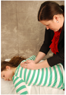
1. Indirect Myofascial Release of Cervico-thoracic Inlet
Physician places thumbs on the 1st rib near the cervico-thoracic junction. Evaluate freedom of movement in rotation and sidebending. Gently apply pressure with thumbs into the direction of freedom of motion in rotation and sidebending until tissue relaxes or motion is less asymmetric.
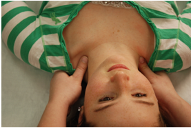 |
Figure 1A. Indirect Myofascial Release of Cervico-thoracic Inlet-Initial Position:
The physician places thumbs on the first rib near the cervico-thoracic junction. |
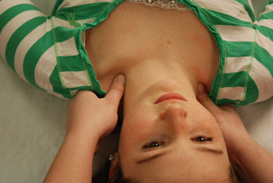 |
Figure 1B. Indirect Myofascial Release of Cervico-thoracic Second Position:
The physician then evaluates freedom of movement in rotation and sidebending. |
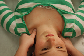 |
Figure 1C. Indirect Myofascial Release
of Cervico-thoracic Inlet-Final Position:
The physician will gently apply pressure with thumbs into the direction of freedom of motion in rotation and sidebending until tissue relaxes or motion is less asymmetric. |
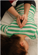
2. Prone Articulatory Technique
Treatment should address areas of greatest restriction. Articulation or “popping” sounds generated by a joint going through motion of secondary areas are of little clinical significance. Patient positioned prone with pillow under his/her chest. Physician positions table to be able to exert a moderate thrust on the posterior transverse process as patient relaxes fully in nearly complete expiration.(14)
3.
Supine Articulatory Technique
This treatment should address lymphatics and restricted areas of respiration. Patient is supine with pillow under the upper dorsal region. Using arms as levers and patient’s inspiration, raise the anterior ends of the upper ribs, moving to cervical and axillary regions. Special attention paid to the first ribs and clavicle.(12, 14)
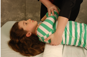 |
Figure 3A. Supine Articulatory
Technique - Initial Position: Patient is supine with pillow under the upper dorsal region. |
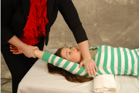 |
Figure 3B. Supine Articulatory
Technique - Final Position: Using arms as levers and patient’s inspiration, raise the anterior ends of the upper ribs, moving to cervical and axillary regions. |
4.
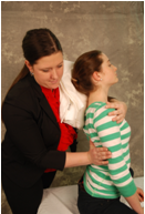
Seated Hyperextension Technique
 |
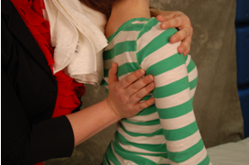 |
Figure 4A |
Figure 4B |
Figure 4A. Seated Hyperextension Technique - Initial Position: Patient sits on stool, table or bed facing away from physician. Patient rests occiput on physician’s shoulder. Physician places one arm across patient’s chest to position patient and the other hand on the ventral side to adjust restrictions. Patient should be relaxed into the physician as the physician neutralizes the spine. |
| Figure 4B. Seated Hyperextension Technique- Final Position: The arm across the chest adds slight traction, rotation, and lateral flexion as hand on ventral side adds thrust anteriorly on the posterior transverse process of restriction.(14) |
5.
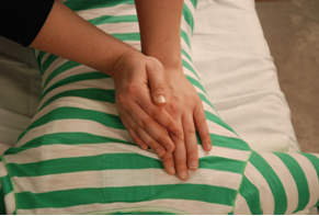
Supine or Seated Thoracic/Abdominal Diaphragm Technique
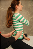
This treatment is aimed at treating the thoracolumbar junction and thoracic diaphragm for improved diaphragm movement. Patient is supine or seated.
 |
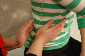 |
Figure 5A |
Figure 5B |
| Figure 5A and Figure 5B Supine or Seated Thoracic/Abdominal Diaphragm Technique: Physician places one hand around the lower torso to contact the tip of the 12th rib. Other hand is place on same area on anterior side. Anterolateral traction is placed on the posterior aspect of the 12th rib. Meanwhile anterior aspect of physician’s hand is moving the diaphragm superiorly and either laterally or medially. Treatment is completed once the thorax moves equally with respiration.(19) |
6.
Additional Treatments to address LRTI
To improve diaphragmatic movement by addressing the Phrenic nerve, treat C3-C5. To decrease lymphatic congestion, treat pelvic diaphragm and thoracic or pedal pumps. To improve parasympathetic effects via the Vagus nerve, treat the OA, AA, and cranium. To decrease sympathetic effects, treat Chapman Reflexes.
Conclusion The Osteopathic approach to treating pediatric patients with lower respiratory tract infections involves the understanding of risk factors, epidemiology, pathogenesis, physical examination finding and common musculoskeletal, lymphatic and autonomic changes in a LRTI. In this article there are high yield OMT techniques that can be readily used in outpatient and inpatient settings that compliment antibiotics and supportive care.
References:
- Rudan I, Boschi-Pinto C, Biloglav Z, Mulholland K, Campbell H. Epidemiology and etiology of childhood pneumonia. Bull World Health Organ. 2008;86(5):408.
- Harris M, Clark J, Coote N, Fletcher P, Harnden A, McKean M, Thomson A, British Thoracic Society Standards of Care Committee. British Thoracic Society guidelines for the management of community acquired pneumonia in children: update 2011.Thorax. 2011;66 Suppl 2:ii1
- Stuckey-Schrock K, Hayes BL, George CM.“Community-Acquired Pneumonia in Children” American Academy of Family Physicians. 2011; Vol 86 (7): 661-667.
- Taylor JA, Del Beccaro M, Done S, Winters W. Establishing clinically relevant standards for tachypnea in febrile children younger than 2 years. Arch Pediatr Adolesc Med. 1995; 149 (3): 283-287.
- Margolis P, Gadomski A. The rational clinical examination. Does this infant have pneumonia? JAMA 1998; 279:308.
- Michelow IC, Olsen K, Lozano J, et al. Epidemiology and clinical characteristics of community-acquired pneumonia in hospitalized children. Pediatrics. 2004; 113 (4): 701-707.
- Virkki R, Juven T, et al. Differentiation of bacterial and viral pneumonia in children. Thorax. 2002; 57 (5): 438-441.
- Harper SA, Bradley JS, Englund JA, et al. Seasonal influenza in adults and children-diagnosis, treatment, chemoprophylaxis, and institutional outbreak management: clinical practice guidelines of the Infectious Diseases Society of America. Clin Infect Dis. 2009; 48 (8): 1003-1032.
- Alberta Clinical Practice Guidelines Steering Committee. Guideline for the diagnosis and management of community acquired pneumonia: pediatric. 2008 update.
- Cincinnati Children’s Hospital Medical Center. Evidence-based care guideline. Community acquired pneumonia in children 60 days through 17 years of age.
- British Thoracic Society Standards of Care Committee. British Thoracic Society guidelines for the management of community acquired pneumonia in childhood. Thorax. 2002; 57 (suppl 1); i1-i24.
- Kuchera, M, Kuchara W. Osteopathic Consideration in Systemic Dysfunction- 2nd edition. 1994 Pg 33-50.
- Chila, AG. Foundations of Osteopathic Medicine-3rd edition. 2011. Lippincott, Williams, and Wilkins.
- Selected writings of Carl Philip McConnell, D.O. 1994 pg 94-9
- Hayes MH, Bezilla TA. Incidence of Iatrogenesis Assoicated with Osteopathic Manipulative treatment of pediatric patients. JAOA. October 2006. Vol 106. No. 10.
- Noll DR, Shores J, Bryman PN, Masterson EV. Adjunctive osteopathic manipulative treatment in the elderly hospitalized with pneumonia: a pilot study. J Am Osteopath Assoc. 1999;99:143-146,151-152
- Noll DR, Shores JH, Gamber RG, Herron KM, Swift J Jr. Benefits of osteopathic manipulative treatment for hospitalized elderly patients with pneumonia. J Am Osteopath Assoc. 2000;100:776-782.
- Noll DR, Degenhardt BF, Fossum C, Hensel, K. Clinical and research protocol for osteopathic treatment of elderly patients with pneumonia. 2008; 108: 508-516.
- Carreiro J. Pediatric Manual Medicine: An Osteopathic Approach. 2009; Pg109. Elsevier.
|