Vitamin D and Bone Health in Perspective
Martha Blackford, PharmD
Michael D. Reed, PharmD, FCCP, FCP
Division of Clinical Pharmacology
Department of Pediatrics and the Rebecca D. Considine Research Institute
Akron Children's Hospital
Akron, Ohio
Introduction
Bone is responsible for the human form, the form of all vertebrate mammals and its health is necessary for optimal body physiology. The need for bone health is unquestionable particularly when one considers that effective bone growth is responsible for determining an individual’s adult height and body mass. A number of factors including complex hormonal axis are responsible for bone growth and the maintenance of bone homeostasis. Of the factors we can more easily control, i.e., exercise, body weight and nutrition, the key mineral components in bone nutrition are adequate vitamin D (Vit D), calcium and phosphate. This review with focus on the important interactions between Vit D, calcium and phosphate and provide the foundations for current recommendations for proper calcium, phosphate and particularly Vit D across the age spectrum.
Bone Physiology – an Overview
Bone is the framework providing structural support and ability for movement of the vertrabrate body. It is a metabolically active connective tissue providing numerous, important physiologic functions including the links for muscle attachment, protection of vital organs, serves as a reservoir for minerals and growth factors, is important to systemic mineral and acid-base homeostasis and is the site for hematopoiesis.(1) Bone affords eventual height and influences overall body mass - it is a dynamic structure continuously remodeling throughout life. In pediatrics, we most often discuss/focus on the growth plate (the physis), a structure that clearly differentiates growing bone during the “pediatric years” and adult bone. Anatomically, the growth plate is located beneath the epiphysis and above the metaphysis. In newborns, the growth plate is essentially absent developing sometime between 12 and 24 months of age.(2) The cells of the growth plate initially convert to rapidly reproducing chrondrocytes that eventually add to height by replicating in a longitudinal manner. Skeletal maturation, on average, is complete in females by 15 to 15.5 years of age while skeletal maturation occurs later in males and is generally complete by 16.5 to 17 years of age.(2)
Bone is very well vascularized serving to maintain needed nutrition for growth. Poor nutrition, disease and/or injury to this vascular network can negatively impact the growing cells and depending upon the magnitude and duration of vascular disruption, damage to future longitudinal growth can occur.(2) Adequate nutrition for optimal bone health involves an adequate for age balanced diet comprised of protein, carbohydrates and lipids with sufficient calories for appropriate anabolism. Minerals and trace metals are also essential for bone stiffness with the primary minerals being calcium and phosphate. Calcium and phosphate are transformed into solid (crystalline) calcium phosphate within the organic matrix of bone.(1) Approximately 90% of bone is comprised of the bone matrix with ~ 65% of the matrix comprising inorganic or mineral matrix, 20% organic matrix and <15% lipids and water. The organic matrix is secreted by osteoblasts and is predominately type I collagen.(1) The osteoprogenitor bone cells are the osteoblasts and these tightly bound cells line the surfaces of bone. Osteoblast activity is marked by alkaline phosphatase (AlkPhos)(1) release which explains the higher AlkPhos concentrations in blood during childhood as compared to adults. The activated osteoblasts, the osteocyte ,maintains the boney matrix and mediates calcium homeostasis.(1) Maintaining bone homeostasis are the osteoclasts, the bone-resportive cells. Multiple endogenous and exogenous factors regulate osteoclast formation and activity including osteoprotegerin, certain interleukins (primarily 1 and 6), macrophage colony-stimulating factor, parathyroid hormone, calcitonin and the focus of this review, 1,25-dihydroxyvitamin D, the active vitamin D analogue.(1)
Vitamin D Metabolism & Clinical Effects
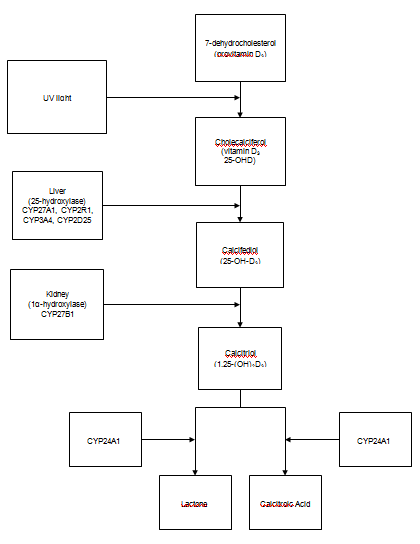
The metabolic disposition of Vit D is shown in the figure.(3) Understanding the metabolic disposition of Vit D is essential to our understanding the optimal means for supplementation, when needed. Vit D is naturally derived from the conversion of 7-dehydrocholesterol in the skin to pre-vitamin D3 (3). This process occurs when the skin is exposed to certain wavelengths of ultra-violet light, the UVB light at 290-315nm; these same wavelengths will also degrade excess pre-vitamin D and Vit D3 thus naturally preventing the risk of intoxication. Pre-vitamin D3 is released into the blood stream and is rapidly converted to vitamin D3 (cholecalciferol) which is further metabolized by vitamin D-25-hydroxylase in the liver, forming the inactive metabolite 25-OHD. The final conversion occurs in the kidney primarily via 25-hydroxyvitamin D-1-α-hydroxylase (also referred to as CYP2R1) which forms the active metabolite 1,25- dihydroxyvitamin D (1,25-(OH)2D). The production of the active metabolite is regulated by serum concentrations of calcium, phosphorous, and parathyroid hormone. Both 25-OHD and 1,25-(OH)2D are inactivated via 25-hydroxyvitamin D-24-hydroxylase (also referred to as CYP24A1) which forms water-soluble calcitroic acid, see Figure 3-5.) (For a comprehensive review of vitamin D metabolism, see references 3 and 4.) To date, few therapeutic compounds have been shown to modulate 25-hydroxyvitamin D-24-hydroxylase (CYP24A1) activity (e.g., enzyme inducers or inhibitors). However, the concentration of the active metabolite 1,25-(OH)2D, induces 25-hydroxyvitamin D-24-hydroxylase (CYP24A1) activity – the greater the active 1,25-(OH)2D concentration the greater the activity (induction) of 25-hydroxyvitamin D-24-hydroxylase (CYP24A1).(3)

Figure. Metabolic disposition of 7-dehydrocholesterol present in the skin to the active form of Vitamin D, calcitrol or 1,25-(OH)2D3. Active Vit D is metabolized primarily by CYP24A1 to inactive metabolites.
Adequate Vit D body stores are important since it affects the extent of dietary calcium and phosphorous absorption. Activated Vit D binds to the vitamin D receptor (VDR) which is part of the superfamily of nuclear hormone receptors.(6) Once 1,25-(OH)2D binds to the receptor, it heterodimerizes with the retinoid-X receptor which then results in regulation of gene transcription after the complex interacts with DNA sequences on these genes; active 1,25-(OH)2D affects numerous functions throughout the body. In the small intestine, the 1,25-(OH)2D-liganded VDR regulates the expression of the epithelial calcium channels and also calcium transporters necessary for calcium to reach systemic circulation.(3,6) In vitamin D deficient states, calcium transport still occurs through passive diffusion however this accounts for only ~10-15% of calcium absorption.(7) Non-skeletal functions of 1,25-(OH)2D have been identified due to the expression of the VDR in various tissues including immune cells, brain, breast, prostate, and colon tissues and/or expression of the 25-hydroxyvitamin D-1-α-hydroxylase.
Historically, Vit D deficiency has been associated with rickets which fortunately has become an unusual diagnosis given the improvements in nutrition over the past century. Unfortunately rickets remains an important disease in infants and children living in less developed countries; Vit D deficiency is still present despite advances in medicine, however patients may present with symptoms other than rickets.(8) Vit D is essential in maintaining the balance between serum calcium, phosphate, and PTH which all factor into appropriate bone mineralization.(7) Pathologic fractures and non-specific musculoskeletal pain or weakness, particularly in adolescent patients who would not be expected to present with classic symptoms of rickets since growth plates fusion is nearing completion,(7) can be an indication of Vit D deficiency; deficiency is also likely to affect a patient’s recovery following orthopedic surgery.(8) Another complication that frequently occurs in obese patients is slipped upper femoral epiphysis, which has not been specifically linked to Vit D deficiency in children but may be suspected.(8) In 64 obese adolescent Polish patients, 86% were found to be insufficient or deficient (defined as 25-OHD <20ng/mL) and their 25-OHD levels were only positively correlated with HDL concentrations and the summer season (June through November).(9) There was a negative correlation between 25-OHD and fasting insulin concentrations and insulin resistance in these obese patients which, if a stronger link is identified, may have an impact on the development of type 2 diabetes mellitus.(9)
Vitamin D
In the past few years, Vit D has become a popular compound. Featured in many periodicals, on the internet and in television advice/talk/medical shows, Vit D supplementation (often with calcium) is frequently touted. The results of this increasing interest in Vit D as a “magic substrate” for many non-skeletal diseases involving many areas of medicine can be confusing, particularly as it pertains to the myriad of supplements available and what seems to be every changing dosing recommendations. Further complicating the definition of optimal Vit D dosing and/or appropriate Vit D body stores is the continued controversy regarding how often to monitor serum concentrations. Consensus suggests that the best approach to monitoring a patient’s Vit D status and/or response to supplementation is monitoring the serum 25-hydroxyvitamin D (25-OHD) concentration (see Figure) and currently accepted serum concentration guidelines is shown on Table 1. Serious Vit D deficiency associated with rickets and osteomalacia is considered when serum 25-OHD concentrations fall below 12 ng/ml (< 30 nmol/L). Vit D deficiency has been defined as serum 25-OHD concentrations <20 ng/mL (50 nmol/L) while patients with serum concentrations between 21-29 ng/mL (52-72nmol/L) are considered insufficient.(3-5,11) Ideally, higher 25-OHD concentrations may be desired but can be difficult to achieve thus concentrations >30ng/mL (75nmol/L) are considered sufficient by most organizations, however serum concentrations >50 ng/ml may not provide additional benefit and/or be associated with adverse effects (see Table 1). Even so, Vit D deficiency is extremely common with incidence reports varying between 20-100% depending on the patient population.(3,5,10) A national United States sampling of pediatric patients aged one to 11 years, found the average serum 25-OHD concentration to be 70nmol/L and the prevalence of serum concentrations <75nmol/L was 65%.(11) Confusion regarding targeted serum Vit D concentrations has been augmented over the years by differences in laboratory assay methodology. This confusion has largely resolved with better, but not absolute, consensus on standards for the quantitation of serum 25-OHD concentration. Any questions regarding possible variability in serum 25-OHD concentration should be directed to your laboratory directory. Also, if you are seeing a patient who has a history of fluctuating serum 25-OHD concentrations and or concentrations discordant from one you may have recently obtained check with the laboratories responsible for the analysis in addition to your detailed medication, vitamin and herbal product history. Note that many products our patients may be self-prescribing may contain various forms of Vit D that is not easily identifiable to the consumer. For example, product labels may denote their Vit D content as ergocalciferol (Vit D2) or cholecalciferol (Vit D3), names that may not be easily interpreted by the patient/consumer.
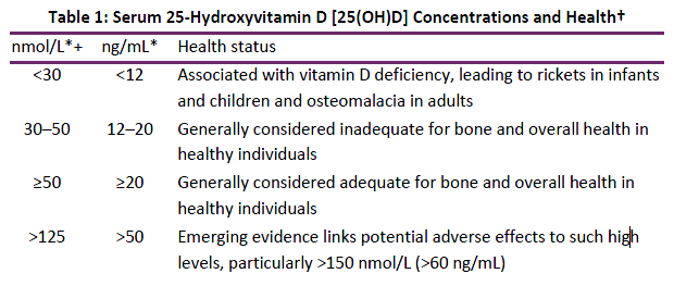
†Adapted from National Institutes of Health Office of Dietary Supplements (ods.od.nih.gov/factsheets/VitaminD-HealthProfessional/)
* Serum concentrations of 25(OH)D are reported in both nanomoles per liter (nmol/L) and nanograms per milliliter (ng/mL).
+ 1 nmol/L = 0.4 ng/mL
Vitamin D Supplements
Dietary vitamin D either comes from an animal or plant source in the form of cholecalciferol (Vit D3) or ergocalciferol (Vit D2), respectively (see Table 2 for dietary sources of vitamin D). Vit D2 is metabolized via the same pathway as Vit D3 (see Figure) however, it has been debated if one form is more effective than the other. Armas et al studied the effects of vitamin D supplementation in healthy adults who were randomized to receive no supplement, 50,000 units of ergocalciferol or 50,000 units of cholecalciferol(12) as a one-time dose and serum concentrations of 25-OHD were monitored periodically over the course of 28 days. Within the first three days post-dose, serum 25-OHD concentrations increased for those receiving supplements compared to the control group and there was no significant difference between the type of vitamin D supplement. However, for those receiving ergocalciferol (Vit D2), 25-OHD, concentrations returned to baseline concentrations by day 14 compared to the cholecalciferol (Vit D3) group whose serum concentrations were still above their baseline at day
28.
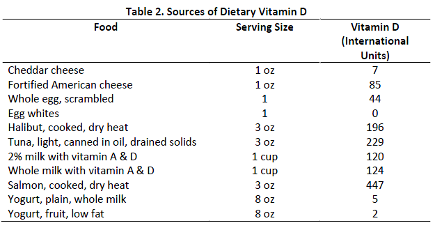
* Adapted from the USDA National Nutrient Database for Standard Reference (www.ars.usda.gov)
Trang et al(13) randomized 34 healthy adults to receive 4000 units/day of Vit D2 or Vit D3 ; two additional groups were included in the study, one received 4000 units of Vit D3 in order to study the changes on serum concentration and the other group acted as a control group with no supplementation. Vitamin supplementation significantly increased the serum concentrations from baseline however, there was also a statistically significant difference in the change from baseline between the two forms of vitamin D, 13.7nmol/L Vit D2 versus 23.3nmol/L Vit D3 [mean difference 9.6nmol/L, (95% CI 1.4-17.8nmol/L)]. It was also noted that patients with a higher baseline vitamin D concentration did not have as great of an increase compared to those with lower baseline concentrations. The authors concluded from this study that Vit D3 was more effective at raising serum concentrations compared to Vit D2 on a per mole basis and should be the preferred supplement.(13) This same rationale has been postulated by others and that Vit D3 possesses a higher affinity for the VDR than Vit D2.(14) Other species have exhibited a difference in response to the type of Vit D used for supplementation, ergocalciferol was more effective for rats while cholecalciferol was more effective in monkeys.(14) Thus, it seems plausible that one form of Vit D may be more effective than another form in humans. Holick and colleagues(15) studied the effects of daily Vit D supplementation for 11 weeks in 68 healthy adults; the patients were randomized to receive either placebo, 1000 units of ergocalciferol (Vit D2), 1000 units of cholecalciferol (Vit D3), or 500 units of both Vit D2 and D3; serum 25-OHD2 and 25-OHD3 concentrations were obtained at baseline and weekly throughout the study. At baseline, 87% of the patients were Vit D insufficient while 60% were deficient. Vitamin D concentrations increased across all three groups (mean change from baseline ranged from 8.2-9.9ng/mL) receiving supplementation with no statistically significant difference between the groups; the primary difference was reflected in the change in 25-OHD2 or 25-OHD3 which corresponded to the form of vitamin D supplemented. The authors also pointed out in this study that Vit D concentrations reached a plateau by week six and the mean steady state concentration of Vit D did not exceed 30ng/mL.
Foods (typically milk, bread, and orange juice) have been fortified with Vit D, either Vits D2 or D3, since the 1930’s. Biancuzzo et al(16) compared the effects of 1000 units of Vit D (both D2 and D3 were studied) supplementation or placebo in either orange juice or a capsule over an 11 week period; 105 otherwise healthy adults were randomized, irrespective of vitamin D status. At baseline, 85% of the patients were Vit D insufficient or deficient (average baseline 25-OHD 17.6 ± 6.4ng/ml); by the end of the study, Vit D serum concentrations increased on average by 9.3-12.8ng/mL across the treatment groups although a plateau was noted in the serum concentrations by week five. Bioavailability was found to be similar between the Vit D in the orange juice and capsules and there was no significant difference between the two forms of Vit D on their effects on serum concentrations.
In a pediatric study, 40 infants and toddlers, ages eight to 24 months with Vit D deficiency (25-OHD ≤20ng/mL), were randomized to receive 2,000 units/day of Vit D2, 50,000 units/week of Vit D2, or 2,000 units/day of Vit D3 for a total of six weeks. (17) All regimens significantly increased serum vitamin D concentrations from baseline with the post median concentration of 36ng/mL (25th-75th percentile range 23-70ng/ml); only three patients did not have serum 25-OHD concentrations that exceeded 20ng/ml however, poor compliance was suspected in each case. The authors concluded from their study that both Vit D2 and D3 supplementation were effective and that weekly doses of Vit D2 were tolerated and effective, as well.
Critical assessment of these publications suggests that the reason(s) for some of the differences observed in results trying to identify which form of Vit D is more effective may be due to the dosing regimen employed, patient population, and the patient’s baseline Vit D status. Daily dosing of either Vit D does not appear to cause a significant difference in serum concentrations however larger, daily or single-doses may have an effect(13,12) although this was not demonstrated in all studies.(15) Even in the study by Trang et al,(13) both forms (Vit D2 or D3) significantly increased serum 25-OHD concentrations and the mean serum 25-OHD for both groups increased to 57.4nmol/L for D2 and 64.6nmol/L for D3, which some groups would consider normal/therapeutic.
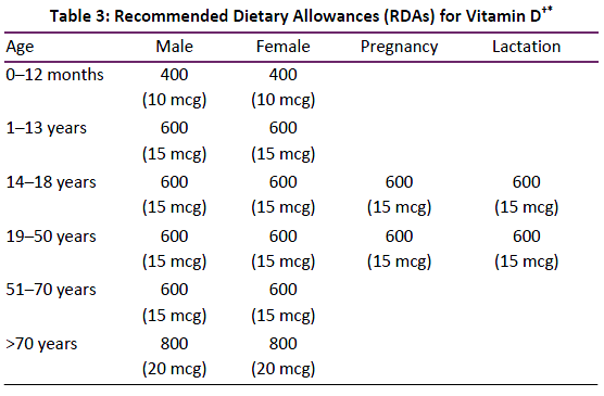
In light of the contrasting findings, the Endocrine Society Clinical Practice Guidelines currently recommends either Vit D2 or D3 as effective sources of Vit D therapy/supplementation.(5) The update in the guidelines for Vit D supplementation in 2011 increased the recommended dosing for most pediatric and adult patients without significant risk factors to 600 units/day (400 units/day if <1 year of age; 800 units/day if >70 years of age). Current recommended dietary allowances (RDAs) for Vit D across the age spectrum for gender and including pregnancy and lactation are shown in Table 3.(5,11) The amounts outlined in Table 3 represent recommended daily Vit D amounts (doses) obtained from food or by Vit D supplementation or both. Examples of commonly used Vit D supplements are shown in Table 4 and the tolerable upper levels of intake in Table 5. If patients are deficient, they may require Vit D supplementation between 1000-2000 units/day to increase serum concentrations to target values, usually >30ng/mL (see Table 1). Some European countries use “stosstherapie” which consists of megadoses of Vit D, 200,00-600,000 units as a one-time dose orally or IM, for Vit D deficient rickets.(7) It is estimated that approximately 100 units of Vit D will increase serum 25-hydroxyvitamin D (25-OHD) concentrations by 1ng/mL.(15)
† Dosage in international units unless otherwise stated
* Adequate Intake adapted from National Institutes of Health Office of Dietary Supplements (ods.od.nih.gov/factsheets/VitaminD-HealthProfessional/); consistent with reference 5.
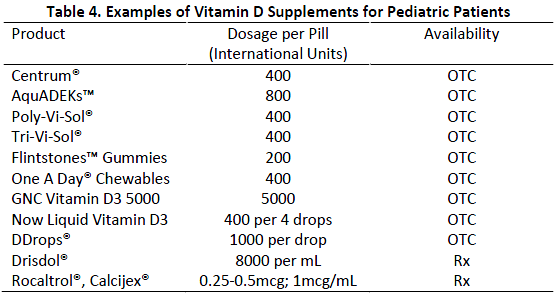
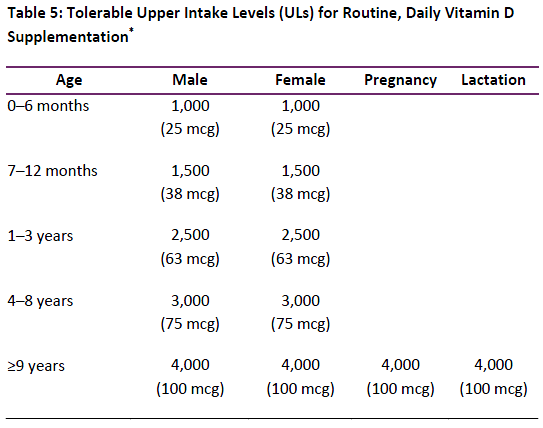
*Dosage in international units unless otherwise stated; National Institutes of Health Office of Dietary Supplements (ods.od.nih.gov/factsheets/VitaminD-HealthProfessional/)
Risk Factors for Deficiency
There are several risk factors for vitamin D deficiency. Patients living at higher latitudes (>35-40o N or S of the equator), elderly, those with malabsorption of fat-soluble vitamins (e.g. cystic fibrosis, inflammatory bowel disease, Crohn’s disease), darker skin pigmentation, minimal skin exposure to sunlight, and/or those receiving certain medications including, glucocorticoids, antifungals (e.g. ketoconazole), possible select anticonvulsant medications and AIDs patients receiving AIDS treatment medications are at higher risk.(5,7) Specific comments regarding drug-associated/drug-induced Vit D deficiency and/or bone disease is addressed below. The family’s routine diet should also be evaluated regarding balanced nutrients, vitamins, minerals and trace metals, particularly for select vegan diets.
Darker skin pigmentation and sunscreens reduce the extent of 7-dehydrocholesterol conversion in the skin to pre-Vit D3 (see Figure); in a national sample across the United States, a higher percentage of non-Hispanic black and Mexican American children were found to have serum 25-OHD concentrations <75nmol/L (< 30 ng/ml) compared to non-Hispanic white children, 89%, 77%, and 54% respectively.(10) It has been estimated that 10-15 minutes of direct sun exposure (depending on the correct wavelength of light), generates 10,000-20,000 units of Vit D(18). Thus it has been suggested that ~15 minutes (3-5 times longer if darker skin pigmentation) of unprotected sun exposure between the hours of 10 am to 3 pm is adequate for Vit D production. However, because of the concerns of sun exposure and skin cancer, Vit D supplementation may be the preferred and easier to control option.
Strictly breastfed infants are at risk for Vit D deficiency, especially if the mother was deficient during pregnancy, since Vit D does not cross into the breast milk very well(7); see Table 3). Most formulas for infants are fortified with Vit D which is protective. However, if a product or milk is being used that was not specifically formulated for infants (e.g. goat’s milk, soy milk), the product may not be fortified with Vit D and a clinically important deficiency spectrum may arise (see Table 1. Vit D status should be vigorously evaluated in such patients and if warranted, Vit D supplementation prescribed (see Table 4).
Multiple drugs have been described to interfere with Vit D absorption or possibly stimulate the activity of Vit D metabolizing enzymes leading to drug-induced rickets, osteoporosis, osteomalacia. Over 50 years ago published reports began to incriminate the anticonvulsants as a common class of drugs leading to drug-induced rickets in children. It was initially hypothesized that the anticonvulsants stimulated the metabolism of Vit D (e.g., phenobarbital, phenytoin [Dilantin®], carbamazepime [Tegretol®]) leading to low serum Vit D concentrations and the subsequent development of bone disease. It is important to note that valproic acid (Depakote®), an enzyme inhibitor, has also been incriminated in bone disorders.(19,20) However, persistent study has been unable to confirm or identify a plausible mechanism of why patients receiving anticonvulsants, particularly multiple anticonvulsant medications daily, may develop osteomalacia, but it does not appear to be related to the effects of anticonvulsants on drug metabolizing enzymes. The early findings in pediatric patients most often involved institutionalized patients and their lack of sun exposure may serve as an important etiology – poor diet may also be a contributing factor. Nevertheless and despite a lack of a clear mechanism, many patients receiving multiple anticonvulsant drugs daily will have a low serum Vit D concentration underscoring the importance of checking a Vit D serum concentration in these patients. In contrast to the anticonvulsants, oral glucocorticoid (steroid) therapy (e.g., prednisone, dexamethasone, Medrol) at a minimum dose equivalent of 5 to 7.5 mg prednisone daily for >3 months is at markedly increased risk of developing osteoporosis.(19,21) Chronic steroid use increases bone loss by reducing intestinal calcium absorption, increasing renal calcium excretion as well as the effects of steroid-induced inhibition on osteoblast function, the reduction in estrogen and testosterone production and the stimulation of parathyroid effects.(19,21) Other drugs that may be associated with osteoporosis include the aromatase inhibitors (adjunct drugs used in post-menopausal women with estrogen-receptor positive breast cancer), and certain antidepressant, antipsychotic and anticoagulant (e.g., warfarin [Coumadin®] drugs, the later agents are highly variable in their bone mineral loss effects. Of possible clinical significance in children is the long-term use of loop diuretics (e.g., furosemide [Lasix®]) by increasing renal calcium excretion and concurrently interfering with the diurnal rhythum of serum parathyroid hormone.(19-21) Additionally, long term use of estrogen-based contraceptives (in contrast to progesterone-based, e.g., DepoProvera®) have been shown to clearly lead to demineralization of bone.
Monitoring Serum Vitamin D Concentrations to Guide Therapy
Although 1,25(OH)2D is the active form (see Figure), routine clinical monitoring for Vit D status involves monitoring the serum 25-OHD concentration. Numerous studies have shown that the 25-OHD metabolite provides a better indicator for total body stores of Vit D when compared to the serum 1,25-(OH)2D concentration which can vary depending on renal function, it is present in the systemic circulation at concentrations ~1000 times lower than 25-OHD, and its production is regulated by several different hormones. The half-life of 1,25-OHD, the active Vit D moiety is significantly shorter than 25-OHD (see Figure), four to 15 hours compared to two to three weeks respectively.(5,22,23) The storage of lipophilic Vit D2 and D3 within adipose tissue accounts for the slow body turnover, approximately two months, for these precursors (see Figure) and also explains the delay in development of low serum concentrations and deficiency-associated bone disease with Vit D deficient consumption. In addition, there appears to be a poor correlation between rickets and the serum concentration since 1,25-(OH)2D concentrations may be low, normal, or high in these patients.(5,7) Testing for 1,25-(OH)2D may be necessary depending on the patient’s underlying condition or suspected condition such as marked/chronic renal disease or chronic granuloma-forming disorders but should not be utilized to routinely assess a patient’s Vit D status. As noted above, most all correlations of serum Vit D concentration with health effect is linked to serum 25-OHD concentration (see Table 1).
Seasonal variations occur naturally in human 25-OHD serum concentrations relative to the degree of sun exposure, skin pigmentation, latitude, and time of day.(4) Patients living in warmer climates are less likely to have seasonal variations although they may still be insufficient or deficient depending on their use of sun screens and degree of skin pigmentation. Because Vit D is fat-soluble, it is stored in the fat tissue therefore during periods of decreased sun exposure/ Vit D production, serum concentrations can be maintained via release from fat stores.
To date, guidelines have remained vague as to the suggested frequency of monitoring. Healthcare providers should take into consideration what season it is being checked and a patient’s risk factors for deficiency. Yearly monitoring may be appropriate if the patient is vitamin D sufficient while patients being treated for insufficiency or deficiency may need for frequent monitoring, especially until they reach their goal.
Bone Mineral Density
Bone mineral density (BMD) can be assessed by a dual-energy X-ray absorptioimetry (DXA) scan in which measurements are generally obtained from the spine, hip, or whole body. For children, the interpretation should reference a Z-score while in adults the T-score is utilized. Children with lower BMD are only considered to have osteoporosis if there has been a pathologic fracture of a long bone in the lower extremities, vertebral compression fracture, or two fractures in a long bone of the upper extremities(24). It remains unclear at this time how pediatric patients with low BMD should be managed. A Cochrane Review on the effects of Vit D supplementation on BMD in children found it may have clinically relevant effects on peak bone mass if the child is Vit D deficient (25). However in children who were not deficient, the review did not find supportive evidence for supplementation and improved bone health.
Because of the positioning required for a DXA scan, accurate results can be difficult, if not impossible, to obtain in children who are unable to remain still or they have contractures, such as with patients with cerebral palsy. A newer technique, in which the distal femur is scanned instead of the lateral femur, has been developed and allows for more comfortable positioning of the patient.(26) An additional benefit of this technique is that the scan involves a region where fractures are more likely to occur in patients with cerebral palsy. In rare cases where such analysis is deemed essential to the diagnosis and treatment, the younger pediatric patient may require procedural sedation for an effective study.
Calcium and Other Substrates
The focus of this review has been Vit D and its congeners reflecting the renewed, heightened interest in this compound. Our main focus has been Vit D effects relative to bone health. However, as noted in our review, Vit D’s impact on optimal bone health is related to its regulation of plasma calcium and phosphate homeostasis and by doing so, maintaining bone mineral homeostasis. For optimal bone health, adequate nutritional intake is required, including calcium – Vit D simply facilitates intestinal calcium absorption and maintains systemic homeostasis.(27-29) As would be expected, daily calcium requirements are age dependent - current recommended daily calcium requirements relative to a patients age is shown in Table 6. Important calcium containing foods and their approximate content are shown in Table 7. As noted above in calculating an individual’s daily Vit D requirements, one needs to assess and account for all possible sources of calcium (e.g., normal foods, daily vitamin and/or other supplements) in calculating the patients added daily calcium need. Dieticians may provide assistance in assessing and developing the overall dietary plan for a patient, especially if a patient has certain food restrictions or special dietary needs. Once the plan has been implemented a plasma calcium (total and “ionized”) concentration should be checked to determine adequacy of the plan.
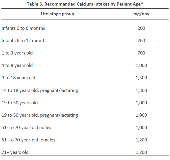 *Adapted from the National Institute of Arthritis & Musculoskeletal and Skin Diseases and National Institute of Health: Calcium and Vitamin D (http://www.niams.nih.gov/Health_Info/Bone/Bone_Health/Nutrition/) *Adapted from the National Institute of Arthritis & Musculoskeletal and Skin Diseases and National Institute of Health: Calcium and Vitamin D (http://www.niams.nih.gov/Health_Info/Bone/Bone_Health/Nutrition/)
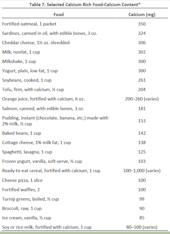
*Adapted from the National Institute of Arthritis & Musculoskeletal and Skin Diseases and National Institute of Health: Calcium and Vitamin D (http://www.niams.nih.gov/Health_Info/Bone/Bone_Health/Nutrition/)
Future Considerations
As addressed throughout this review, there are several aspects of Vit D supplementation that remain unclear. Optimal supplementation doses and even which form of Vit D is best, continue to be debated. Patients who are Vit D insufficient or deficient can easily be treated with supplements but for patients with low BMD, it is unclear what the best approach should be or long term consequences. It is very likely that genetic variants of the VDR, metabolizing enzymes, or other components of Vit D pathway affect a patient’s response to Vit D however the clinically relevant variants have yet to be determined.(30,31)
References:
- Buck DW, Dumanian GA. Bone biology and physiology: Part I. The fundamentals. Plast Reconstr Surg 2012;129:1314-1320
- Weiner DS, ed. Pediatric orthopedics for primary care physicians. 2nd edition, Cambridge Univ Press;2004:1-8
- Henry HL. Regulation of vitamin D metabolism. Clin Endo Metab 2011;25:531-541
- Holick MF. Vitamin D deficiency. N Engl J Med 2007; 357: 266-81.
- Holick MF, Binkley NC, Bischoff-Ferrari HA, Gordon CM, Hanley DA, Heaney RP, Murad MH, Weaver CM. Evaluation, treatment, and prevention of vitamin d deficiency: an Endocrine Society Clinical Practice Guideline. J Clin Endocrinol Metab 2011; 96: 1911-30.
- St-Arnaud R, Naja RP. Vitamin d metabolism, cartilage, and bone fracture repair. Mol Cell Endocrinol 2011; 347: 48-54.
- Pettifor JM, Prentice A. The role of vitamin D in paediatric bone health. Best Prac Res Clin Endocrinol Metab 2011; 25: 573-84.
- Clarke N, Page JE. Vitamin d deficiency: a paediatric orthopaedic perspective. Curr Opin Pediatr 2012; 24:46-9.
|