


 |
|||||||||||||||||
Contents The Neuroprotective Benefits of Therapeutic Hypothermia in Pediatric Near-Drowning with Cardiac Arrest: A Case Report _________ Trisomy 9: A Case Report of Neurological _________ Obesity, Type Two Diabetes Mellitus, Asthma All _________ Osteogenesis Imperfecta: A Complicated Diagnosis _________ The Neuroprotective Benefits of Therapeutic Hypothermia in Pediatric Near-Drowning with Cardiac Arrest: A Case Report
Introduction HIE can cause multisystem dysfunction, specifically involving the pulmonary, cardiovascular, renal, and gastrointestinal systems. From a pulmonary standpoint, surfactant wash-out results in respiratory distress syndrome (RDS). Increased capillary permeability causes intrapulmonary shunting with V/Q mismatch, atelectasis, and poor compliance. From a cardiovascular viewpoint, the anoxia damages muscle, creating global dysfunction and arrhythmias. Arrhythmias are worsened by metabolic derangements and electrolyte shifts. If hypoxia is prolonged, cardiac arrest ensues. From a renal point of view, rhabdomyolysis from extensive tissue destruction then leads to acute tubular necrosis (ATN) with varying degrees of oliguria and azotemia. Inappropriate ADH is common in the first 72 hours post-HIE, and the low osmolarity and hyponatremia increase cerebral edema and seizures. From a GI standpoint, luminal ischemia predisposes to necrotizing enterocolitis. Hepatocellular injury results in impaired synthetic function, resulting in hypoglycemia and coagulopathy, often to the extent of disseminated intravascular coagulation (DIC).(5) In hypoxic ischemic encephalopathy, the degree of disability is related to duration of cardiac arrest and secondary damage post resuscitation. Hypoxia for a few minutes can cause neuronal death in primary injury. Additionally, CNS injury is temperature dependent. Fever worsens neurotransmitter and oxygen free radical production, the blood-brain barrier fails, depolarization in the ischemic penumbra is damaged, there is cytoskeletal proteolysis, and impaired recovery of energy metabolism. Therapeutic hypothermia minimizes the secondary injury via different mechanisms. Therapeutic hypothermia decreases metabolism, modulates intracellular signaling and gene expression. These functions control the cascades of oxidative injury, inflammation, and apoptosis during ischemia and reperfusion, resulting in reduced number of neurons lost.
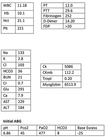
PICU Course An initial EEG reading was diffusely depressed with a slow background. The initial echocardiogram showed grossly normal anatomy with global hypodyskinesia, more pronounced in the left ventricle. The patient’s PICU course was complicated by rhabdomyolysis, hyperglycemia, aspiration pneumonia, ARDS, and cardiogenic shock. All of these complications prolonged his mechanical ventilation, but were aggressively treated and resolved. His repeat echocardiogram was normal for age, and his EKG showed abnormal prolonged QT with low amplitude QRS. His post-EEG, when discontinued after 36 hours, was continuous and attenuated with improvement and showed sleep spindles without seizures. On day 10, the patient was extubated with an arterial blood gas of 7.40/45/107/28/3. The patient’s extubation was prolonged by fluid overload and aspiration pneumonia, secondary from the near-drowning. Once extubated, the patient continued to improve, and his repeat chest x-ray was mostly cleared, with improvement in haziness. On day eleven of the patient’s PICU stay, his physical exam showed significant improvements, revealing PERRLA of the eyes (4mm), good bilateral air exchange with occasional rhonchi, a non-distended and non-tender abdomen with positive bowel sounds, and a cardiovascular exam exhibiting S1, S2 and 2+ peripheral pulses. The patient’s neurological exam revealed hypotonicity, and he was following commands well, but was non-verbal. Inpatient Pediatrics Course Therapeutic Hypothermia Protocol Labs drawn were due to physiologic changes observed under hypothermia at specific intervals illustrated by the adult studies. The physiologic changes seen in the adult studies were noted to affect each organ system, and the timing of the lab draws were based on the adult protocol.(3) For the renal system, decreased creatinine clearance and central redistribution of volume can cause diuresis, dehydration, and electrolyte imbalances. The gastrointestinal system can have increased dysfunction, requiring monitoring of liver function tests. Coagulation may be disturbed by decreased enzyme activity, enhanced fibrinolysis, platelet dysfunction, and a diffuse DIC-like syndrome. HIE is often complicated by diabetes insipidus and SIADH, which is why frequent urinalyses are performed. Labs obtained during the protocol consisted of:
Discussion This case illustrates the potential for therapeutic hypothermia to decrease the References:
_______________ Trisomy 9: A Case Report of Neurological Sequelae and Literature Review Josi Herren, BS Abstract Introduction Case Presentation
|
|||||||||||||||||
Title |
Summary |
Kannan, T. P. et al. Clinical manifestations in trisomy 9 |
Case discussion of fetus ending in normal delivery and surviving until 20 days is reported here detailing the clinical manifestations of the child during the period of survival. The salient clinical features observed were small face, wide fontanel, prominent occiput, micrognathia, low set ears, upslanting palpebral fissures, high arched palate, short sternum, overlapping fingers, limited hip abduction, rocker bottom feet, heart murmurs and also webbed neck, characteristic of this trisomy 9 syndrome. |
Ferreres, J. et al. Pathological findings in the complete trisomy 9 syndrome: three case reports and review of the literature. |
Review of pathological autopsy examination of “complete trisomy 9” without mosaicisms. |
Patil, S. J. et al. Mosaic trisomy 9 presenting with congenital heart disease, facial dysmorphism and pigmentary skin lesions: Intricate issues of genetic counseling. |
A case presentation of mosaic trisomy 9 with typical manifestation (facial dysmorphism, various internal organ malformations and severe phsychomotor retardation) and pigmentary skin lesions |
Kor-Anantakul, O. et al. Prenatal diagnosis of complete trisomy 9: A case report and review of the literature. |
A case report of complete trisomy 9 with prenatal sonographic findings in the second trimester. The combination of sonography and karyotyping from cordocentesis, established this prenatal diagnosis and reports aortopulmonary communication. Along with above, a literature review specifically focusing on prenatal sonographic findings was presented. |
Sutherland, G. R. et al. Partial and complete trisomy 9: delineation of a trisomy 9 syndrome. Human genetics. |
Case presentation of two infants with trisomy involving chromosome 9 are described. One infant had complete trisomy 9, a lethal diagnosis, and the 2nd infant had a karyotype of 47,XX,+der(9),t(7;9) (p22;q32)mat. The included abnormalities were cardiovascular and urogenital systems, dislocation of the hips, knees, cranial suture abnormalities, and early death. |
Bruns, D. (2011). Presenting physical characteristics, medical conditions, and developmental status of long-term survivors with trisomy 9 mosaicism. American Journal of Medical Genetics, Part A, 155(5), 1033-1039. |
Data from the Tracking Rare Incidence Syndromes (TRIS) project offer the largest series to date examining the physical and medical conditions for 14 individuals with Trisomy 9. Results indicated the presence of low set ears, and microcephaly. The developmental status indicated a wide range in functioning level with the conclusions of younger children demonstrating skills approaching their chronological age, and varied as the children got older. |
Feingold M, Atkins L. A case of trisomy 9. J Med Genet. 1973;10:184–7. |
This case report was the first patient with trisomy 9, who survived 28 days. Clinical findings included microcephaly, low set ears, small palpebral fissures, enophthalmos, bulbous nose, micrognathia, congenital heart disease, skeletal abnormalities, small penis, hypoplastic scrotum, and a Dandy-Walker like deformity of the fourth ventricle. |
_______________
Obesity, Type Two Diabetes Mellitus, Asthma
All Present With Prader-Willi Syndrome
A Case Study
Kyle Hazlett
Biological Sciences
Wright State University
Dayton, Ohio
Carl R Backes, DO, FACOP
Professor Pediatrics
Ohio University College of Osteopathic Medicine
Chairman Dept. Pediatrics Doctors Hospital Ohio Health
Abstract:
The United States has a problem with obesity. In the US,18.4% of teenagers are considered obese and the percentage is even higher in adults at 35.1%.(1) Our case study reinforces complications, correlations of obesity with type II diabetes and asthma and it’s effect in a patient with Prader-Willi Syndrome.
Case Presentation:
Patient X is a 13-year-old Indiana female who weighs 103kg. She presented to Kiddie West Pediatrics complaining of wheezing and shortness of breath. Patient X has previously been diagnosed with Prader-Willi Syndrome (PWS). She has morbid obesity, obstructive sleep apnea, type II diabetes, and asthma. (2) Her FEV1 is 68%. Her hemoglobin A1C is 5.7% and her IGFI is 757. Finally her BMI is 34.6.(2)
Methods/Results:
Treatment included inhaled Albuterol treatment, then adding an inhaled steroid, oral antihistamines and continuing Genotropin 12mg and Glucophage 1000mg bid.(2)
Discussion:
Prader-Willi Syndrome is an uncommon genetic birth defect affecting an estimated 1 in 15,000 people. The syndrome is caused by an abnormal chromosome (15 at q11q13). A patient with PWS presents between 6 months and 6 years of age with an insatiable appetite. They are unable to control their eating habits. Eventually they develop obesity and type 2 diabetes mellitus.(3)
Type II diabetes mellitus (TTDM) is presently the most prevalent form of diabetes. “Symptoms of TTDM include frequent urination, excessive thirst, weight loss, extreme hunger, sudden vision changes, tingling or numbness in the hands or feet, fatigue, very dry skin, sores that are slow to heal and frequent infections.”(4) TTDM is also known as insulin-resistant diabetes. With TTDM the body initially produces enough insulin to break down the extra dietary glucose. Eventually, however, the insulin produced does not work and the body cannot maintain a normal blood glucose level.(5) The risk of developing TTDM increases drastically when the individual is overweight. “Almost 90% of people with type 2 diabetes are overweight.”(6)
The CDC makes a clear difference between overweight diagnosing an individual as overweight or obese. It is based on body mass index (BMI).(7) A normal adult BMI has a range of 18.5 to 24.99. Overweight individuals BMI’s must be in between 25 and 29.9. and anyone’s BMI above 30 is obese. According to the CDC “obesity is associated significantly with the development of asthma, worsening asthma symptoms, and poor asthma control.”(8)
A study conducted at the University at Buffalo found a correlation between obesity and asthma. “The study initially focused on the prevalence of asthma between normal weight and obese patients. It then looked at the behavior of asthma-linked genes and how they we altered when an obese person undergoes gastric bypass surgery. With obesity “Four genes are activated with chronic inflammation.”(9) “The activated genes cause mononuclear cells to produce greater amounts of inflammatory factors (interleukin 4, LIGHT and lymphotoxinb receptor) which contributes to allergic inflammation.(9) There are also two more compounds that affect asthma and are more prevalent in obese patients verse normal weight patients. MMP-9 and nitric oxide metabolites (NOM) are asthma related factors at higher concentrations in morbidly obese patients.”(9) After gastric bypass in obese patients there is a significant reduction in asthma related factors (MMP-9 and NOM levels). The expressing asthma-related genes noted changed.(9) Previously, no biological, mechanistic explanation in asthma diagnosis was made other than obesity raises the diaphragm and reduces lung volumes. Now a biologic mechanism explains the connection between Type II diabetes, obesity and asthma. We, as physicians, can now predict a 100% increased prevalence in asthma when associated with morbid obesity and type II diabetes.”(10)
Patient X has been diagnosed with PWS, with her insatiable appetite and morbid weight gain (BMI 34.6). With associated type II diabetes, we can now explain her newly diagnosed moderate persistent asthma. To eliminate multiple medications patient X uses, she is placed on a restricted diet with daily exercise. We anticipate long standing health issues with her chromosomal abnormality (PWS).
References:
- "Obesity and Overweight." Centers for Disease Control and Prevention. Centers for Disease Control and Prevention, 14 May 2014. Web. 24 Aug. 2014.
- Personal Communication: Backes DO, Carl R
- Jones, Kenneth L., MD. "Unusual Brain And/or Neuromuscular Fingdings." Smith's Recognizable Patterns of Human Malformation (Major Problems in Pathology). 5th ed. United States of America: W.B Saunders, 1997. 202-03. Print.
- "Diabetes Mellitus Signs and Symptoms." Diabetes Mellitus Signs and Symptoms. UCSF Medical Center, n.d. Web. 17 Aug. 2014.
- Type 2." American Diabetes Association. N.p., n.d. Web. 16 Aug. 2014.
- "Your Weight and Diabetes." Obesity.org. N.p., 2010. Web. 19 Aug. 2014.
- "Disability and Obesity." Centers for Disease Control and Prevention. Centers for Disease Control and Prevention, 01 Apr. 2014. Web. 19 Aug. 2014.
- "AsthmaStats." Centers for Disease Control and Prevention. Centers for Disease Control and Prevention, 09 Aug. 2013. Web. 20 Aug. 2014.
- University at Buffalo. “Obesity and Asthma: Study finds a link in the genes.” ScienceDaily. ScienceDaily, 18 July 2013.
- Paresh Dandona, Husam Ghanim, Scott V. Monte, Joseph A Caruana, Kelly Green, Sanaa Abuaysheh, Teekam Lohano, Jerome Schentag, Sandeep Dhindsa, Ajay Chaudhuri. Increase in the mediators of asthma in obesity and obesity with type 2 diabetes: Reduction with weight loss. Obesity, 2013; DOI:10.1002/oby.20524

_______________
Osteogenesis Imperfecta: A Complicated Diagnosis With Concerns For Misdiagnosed Physical Abuse
Kayla Crager
Biological Sciences
Wright State University
Dayton, Ohio
Carl R Backes, DO, FACOP
Professor Pediatrics
Ohio University College of Osteopathic Medicine
Chairman Dept. Pediatrics Doctors Hospital Ohio Health
Abstract:
Osteogenesis Imperfecta (OI) is an uncommon disease, causing defective production of collagen.(3) There are different types of OI, causing misconceptions and difficulty in diagnosing the disease initially. These are two case studies of OI infants with different presentations.
Presentation of Case I:
An 8-week-old male presented to Nationwide Children’s Hospital emergency room with a right spiral femur, a tibial fracture, a left sixth rib fracture, and subdural hematoma initially from alleged physical abuse. Pain medication was given and the leg was splinted. There were no other injuries noted. The parents said the babysitter was with the baby prior to the hospital visit. Child services and the police were notified by the emergency room physician. The mother stated there was no family history of bleeding disorders, no history of previous fractures and no history of trauma. Several relatives on father’s side, however, were diagnosed with osteogenesis imperfecta.(1) A genetic evaluation by Nationwide Children’s Hospital confirmed blue sclerae, no bruising or unusual features, and normal muscle tones. The infant failed a hearing test and bone density was low. Genetic testing confirmed a mutation in COL1A1 gene for both patient and biological father confirming the diagnoses of osteogenesis imperfecta type I.(1)
Presentation of Case II:
We present a female patient, who despite a normal pregnancy in the mother, had several fractured bones in utero including but not limited to the tibia, fibula, humerus and several ribs.(1) After birth, blue sclerae and bone deformities were observed. There was no family history of birthing problems, learning defects, osteoporosis or osteopenia. Her diagnosis of OI type II was confirmed after genetic testing showed the COL1A1 mutation.(1)
Laboratory Results of Case I:
A bone density scan showed a result of .225 g/cm2, 2 months after the followup. Vitamin D2 levels were low. The CBC revealed elevated RBCs, hemoglobin, and lymphocytes. Osteocalcin levels were elevated, calcium level was normal. Six months later a second CBC was obtained and showed an elevated platelet count. At that time phosphorous and osteocalcin levels were still elevated. Calcium level was normal.(1)
Laboratory Results of Case II:
A Babygram revealed osteopenia, several fractures and bowing of bones, and an under-developed skull.(1)
Treatments of Case I:
Patient was treated with acetaminophen and pamidronate Disodium infusions Q 3mo.(1)
Treatments of Case II:
The patient was given acetaminophen and IV pamidronate was recommended. Patient was also treated with several osteotomies.(1)
Discussion:
Osteogenesis Imperfecta(OI) is described as an inherited genetic disorder where collagen is not properly produced, causing fractures of the bone, along with several other signs and symptoms.[3] Collagen is a significant part of the body. It provides strength and durability to parts of the body such as the teeth, bone, eyes, and ligaments.(2) The body uses our genes as directions to produce proteins that constitute our cells, tissues, and organs. The gene COL1A1 codes for collagen type 1, which is the type of collagen that is modified in about 90% of all OI cases. Patients with OI types I, II, III, or IV experience a mutation in this COL1A1 gene, which causes the production of type 1 collagen to be altered or insufficient. This in turn affects the different parts of the body, which have collagen as an important part of its structure.(3)
In the United States about 25,000-50,000 are diagnosed with OI.(3) This disease is not common and it does not discriminate against gender or race. OI is genetically inherited and is normally 90% autosomal dominant. In rare cases (10%), OI is inherited as a recessive gene and affects another form of collagen besides type 1.(3) Common findings associated with OI include bone fractures, blue sclerae, respiratory difficulties, bone deformity, short stature, scoliosis, low bone density, trouble hearing and sometimes fractures in utero as well as dentinogenesis imperfect.(3)
There are several different types of OI. Type 1 is the most prevalent and mild.(3) Our first case presented with blue sclerae, bone fractures, low bone density and failed hearing test. The initial visit presented with several fractures including a spiral fracture of the femur, a fractured rib and tibia.(1)
Most fractures in kids less than 18 months of age are from some type of physical mistreatment.(4) The usual bones that are fractured from abuse are the ribs, femur and tibia as well as other long bones. A concerning type of fracture seen in abuse is a spiral fracture, which results from a twisting motion of the bone.(4) An initial examination of our patient suggested the possibility of physical abuse due to the spiral fracture.
The diagnosis of OI was confirmed after genetic testing revealed the mutation of the COL1A1 gene.(1) Genetic testing is conclusive in diagnosing OI.(2) Our patient was designated with a type 1 OI after physicians reviewed his case. The elevated labs included RBCs, hemoglobin, lymphocytes, osteocalcin and phosphorus.(1) This is due to the bone healing from the multiple bone fractures and lack of bone strength and density. Calcium levels remained normal as expected as OI affects the production of collagen not calcium.(3) The Patient’s BMD scan was .225 g/cm2, putting him in the 3rd percentile for infants from birth to one year of age.(1,5) Patients with type I OI have less fractures after puberty and usually are able to live normal lives without disease complications.
However, patients with type II OI have more serious complications.(2) Type II is the severest form of OI, usually resulting in death.(2) Case II presented with several fractures in utero. Broken bones in utero is a common finding in type II OI, and makes it easier to correctly diagnose OI and rule out abuse.(2) The Babygram revealed the infant had osteopenia, bowed limbs, and a skull that was not fully developed.(1) These are also indications of type II OI.(2) One significant difference between this patient is the fracture timing. In OI type II fractures occur in utero.
Summary:
OI is currently incurable, but there are treatment options including pamidronate infusions. This assists in increasing bone thickness and reducing its resorption.(2) Placing intramedullary rods in bones with osteotomies aids in the improvement of the bone deformities as well as prevention of potential fractures. Another treatment is physical therapy to help strengthen bone and surrounding muscles.(2)
Although OI is a rare disease, these cases show the importance of a thorough examination, history and observing radiographic and laboratory data.
Multiple fractures in utero make the diagnosis easier. Fractures after birth, however, especially spiral fractures are considered abuse until disproven.
References:
- Personal Communication, Carl R. Backes, DO
- Ramachandran, Manoj, Pramod Achan, David Jones, and Vinod Panchbhavi. "Osteogenesis Imperfecta ." Osteogenesis Imperfecta. Ed. Harris Gellman. Medscape, 1994-2014. Web. 18 Aug. 2014.
- "Subscribe to TheBreakthrough Newsletter OrE-News." Osteogenesis Imperfecta Foundation. Osteogenesis Imperfecta Foundation, 2014. Web. 18 Aug. 2014.
- Flaherty, E. G., J. M. Perez-Rossello, M. A. Levine, and W. L. Hennrikus. "Evaluating Children With Fractures for Child Physical Abuse." Pediatrics 133.2 (2014): E477-489. Web. 19 Aug. 2014.
- Gallo, Sina, Catherine Vanstone, and Hope Weiler. "Normative Data for Bone Mass in Healthy Term Infants from Birth to 1 Year of Age." Normative Data for Bone Mass in Healthy Term Infants from Birth to 1 Year of Age. Ed. E.M. Lewiecki. Sina Gallo Et Al., 2012. Web. 24 Aug. 2014.